NAD+ (nicotinamide adenine dinucleotide, oxidized form) is a pyridine dinucleotide coenzyme present in all living cells. In the Ever Vital catalog, NAD+ is listed with the formula C21H27N7O14P2, a molecular weight of 663.43 g/mol (anhydrous free acid) and CAS 53-84-9. In laboratory work it appears in two roles: as a defined reagent added to cell-free enzyme assays, and as the endogenous pool that researchers measure in cell and tissue samples.
This article covers how it is handled in both, and the assay considerations that most often decide whether a result can be interpreted. It is written for research planning only. For the biochemistry of the molecule itself, see the NAD+ research overview and the article on NAD+ structure and redox chemistry.
Why is NAD+ a useful reagent in cell-free enzyme assays?
Cell-free systems reduce a biological question to chemistry. NAD+ is a substrate or cofactor for several enzyme families, so it is the defined input in assays for sirtuin deacylation, PARP-catalyzed ADP-ribosylation, CD38 hydrolysis and many dehydrogenases. The investigator sets buffer composition, pH and enzyme concentration, which makes it possible to isolate a single variable and to measure kinetic parameters.
The weakness of a cell-free format is the reverse of its strength. It lacks compartments, transport and competing pathways, so a clean result in a purified system is a statement about that system. Published methods also stress that the nominal amount of a nucleotide stock is not always its real content. Moisture, partial degradation and counter-ion form all change the effective concentration, so careful protocols quantify the stock spectrophotometrically before use, and they report the quantity measured.
How is the NAD+/NADH ratio measured in cell culture?
The ratio of oxidized to reduced coenzyme is a widely used readout of cellular redox and metabolic state. The measurement is simple in concept and demanding in practice, because the two forms are chemically labile under opposite conditions. The oxidized form is stable in acid and degrades in base, while the reduced form is stable in base and degrades in acid.
Published analyses therefore describe extraction conditions matched to the form of interest, rapid quenching of metabolism at harvest, and low temperatures throughout. Detection formats reported in the literature include enzymatic cycling assays, HPLC with absorbance detection and mass spectrometry. Each has a different sensitivity and a different susceptibility to interference. Researchers interpreting a ratio also account for compartments, since mitochondrial and cytosolic pools differ and a whole-cell lysate averages across them. Genetically encoded fluorescent sensors reported in the literature were developed partly to address that limitation.
Which enzymes consume NAD+ in cultured cells?
Three consumer families dominate the in vitro literature. Sirtuins (SIRT1 to SIRT7 in mammals) use NAD+ for protein deacylation and release nicotinamide. PARP enzymes, especially PARP-1, use it to build ADP-ribose polymers at sites of DNA strand breaks. CD38 is an ectoenzyme that hydrolyzes NAD+ to cyclic ADP-ribose and ADP-ribose.
Because all three draw on the same pool, a change in total NAD+ readout is not specific to any one of them. A careful design pairs the pool measurement with pathway-specific controls, such as a PARP inhibitor, a sirtuin inhibitor or a CD38-deficient line, to attribute a change to a consumer. The same logic applies to synthesis: published work describes salvage and de novo routes feeding the pool, and these can be altered independently in culture. Without such controls a pool measurement is descriptive and cannot support a mechanistic claim.
How does the culture setup influence NAD+ experiments?
Culture conditions shape the NAD+ pool as much as any reagent does. Medium composition determines which precursors are available. Serum contributes its own enzymes, including ones that act on extracellular nucleotides. Cell density, passage number and glucose level all change metabolic flux and therefore the coenzyme balance.
This matters for experiments that add a nucleotide to the medium. Extracellular NAD+ is processed by ectoenzymes at the cell surface, and published studies describe breakdown products being taken up rather than the intact molecule crossing the membrane. A change in the intracellular pool after such an addition may therefore reflect precursor handling and not direct uptake. Studies that care about the distinction include controls with the breakdown products and conditions that inhibit the relevant ectoenzymes.
How is NAD+ studied in cellular aging models?
Longevity-oriented in vitro work uses cultured cells in replicative senescence, serially passaged primary cells and cells under chronic low-level stress. Published analyses compare the NAD+ pool, the NAD+/NADH ratio and the activity of consuming enzymes between early-passage and late-passage populations. The questions are descriptive and mechanistic: how does the coenzyme balance change as cells approach senescence, and which pathway accounts for the change?
These models connect to neighboring catalog topics. Glutathione regeneration depends on reducing equivalents from the related cofactor NADPH, which links redox studies to glutathione in in vitro models. Mitochondrial signaling studies involving MOTS-c touch the same metabolic network. Researchers planning an aging-model experiment often measure several layers together rather than one in isolation.
What does the catalog specification say about the material?
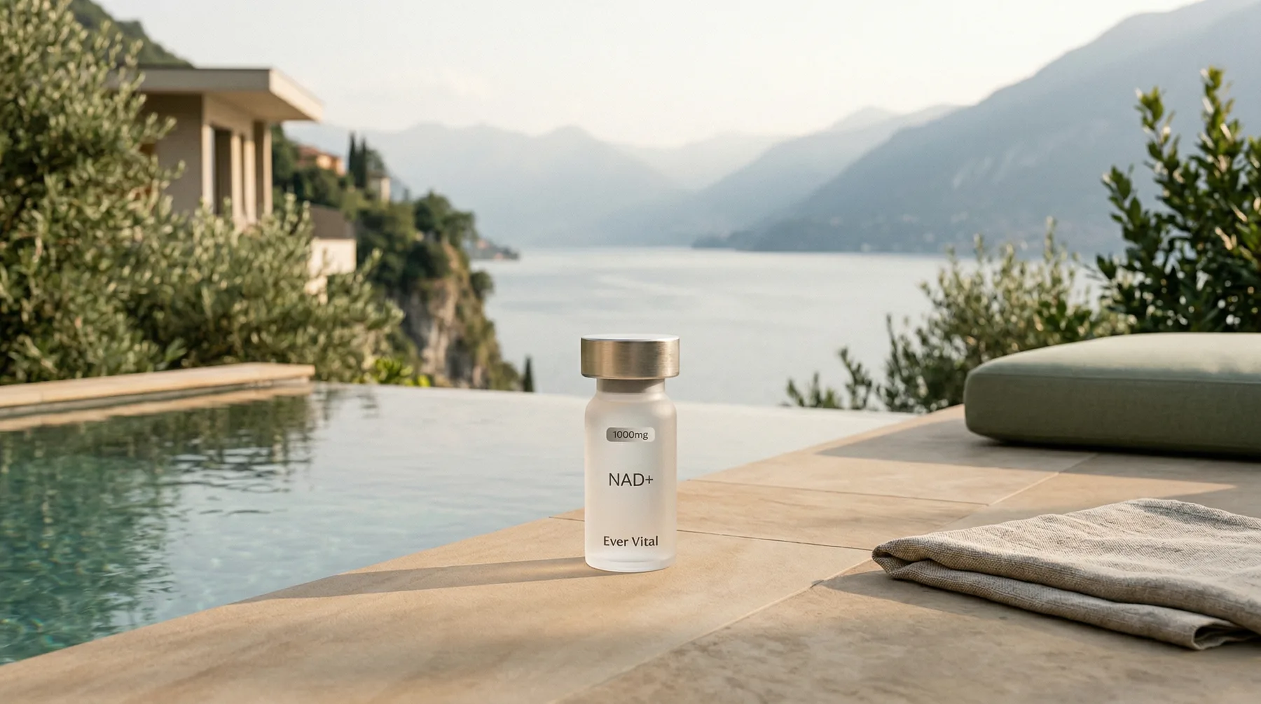
The Ever Vital catalog lists NAD+ at a stated purity of 99% as a lyophilized powder in 500mg and 1000mg sizes, with storage at -20°C. The registry number CAS 53-84-9 and the 663.43 g/mol molecular weight refer to the anhydrous free acid, so hydrated lots carry a higher effective mass. For quantitative work, the practical consequence is that the label mass and the true nucleotide content can differ, which is why a certificate of analysis and an independent spectrophotometric check are part of careful planning.
Identity documentation also protects against confusion with related molecules. NADH, NADP+, nicotinamide mononucleotide and nicotinamide riboside are all discussed in the same literature, carry different formulas and registry numbers, and behave differently in assays. Matching the formula C21H27N7O14P2 and CAS 53-84-9 against the paperwork for the lot in hand keeps a methods section accurate and a result attributable to the right molecule.
What are the common pitfalls in NAD+ in vitro work?
A short list of recurring problems appears across the methods literature.
Degradation during handling is the first. The molecule is sensitive to extreme pH, elevated temperature and prolonged time in solution, so careful protocols keep samples cold, work quickly and check stocks against a fresh standard. Solid material is stored cold and dry according to the supplier specification; this article does not describe solution preparation, which each investigator determines for their own assay.
Specificity is the second. Cycling assays and absorbance readouts can respond to related nucleotides, so orthogonal confirmation by chromatography or mass spectrometry is common practice.
Normalization is the third. Pool size is typically expressed per cell number or per milligram of protein, and the choice changes the interpretation when a condition alters cell size or protein content.
Interpretation is the fourth. Results from isolated enzymes and cultured cells describe those systems. They are not demonstrations of outcomes in organisms, and Ever Vital makes no therapeutic claims about NAD+.
How should an in vitro NAD+ experiment be documented?
A reviewer reading a methods section needs enough detail to repeat the work. The routine below is a documentation habit for research planning, nothing more.
State the system: cell-free enzyme, cell line or primary culture, with passage number and medium.
State the readout: pool, ratio or pathway-specific activity, and the detection method.
State the controls: consumer-pathway inhibitors, breakdown-product controls and a vehicle condition.
Record the material: name, formula, CAS number, lot and certificate of analysis.
This article is provided for informational and research purposes only. The compounds discussed are research chemicals intended for laboratory use only. They are not drugs, supplements or food, and are not intended to diagnose, treat, cure or prevent any disease. Ever Vital does not sell products intended for human use. Researchers are responsible for compliance with all applicable local, state and federal regulations.
Reference
Common questions
In cell-free work NAD+ serves as a defined substrate or cofactor, for example in sirtuin deacetylation, PARP activity and dehydrogenase assays. Because buffer, pH and enzyme concentration are set by the investigator, a single variable can be isolated. Published methods report the actual NAD+ content of the stock used rather than assuming it.
The two forms are labile under opposite conditions: the oxidized form degrades in base and the reduced form degrades in acid. Extraction therefore has to be matched to the form being measured, and delays or temperature changes can shift the apparent ratio. Published methods typically use separate extractions or validated simultaneous protocols.
Three major families are described in the literature: sirtuin deacylases, PARP enzymes active in DNA damage signaling, and CD38, an ectoenzyme that hydrolyzes NAD+. Each draws on the same pool, so a change in total NAD+ readout can reflect any of them, which is why pathway-specific controls are standard.
No. In vitro models describe the behavior of a defined system, such as an isolated enzyme or a cultured cell line. They are useful for mechanism and for comparing conditions, but they do not demonstrate outcomes in organisms, and Ever Vital makes no therapeutic claims about NAD+. Interpretation stays within the model.
Join the list.
Restock alerts, subscriber discounts, and first access to new compound drops.